
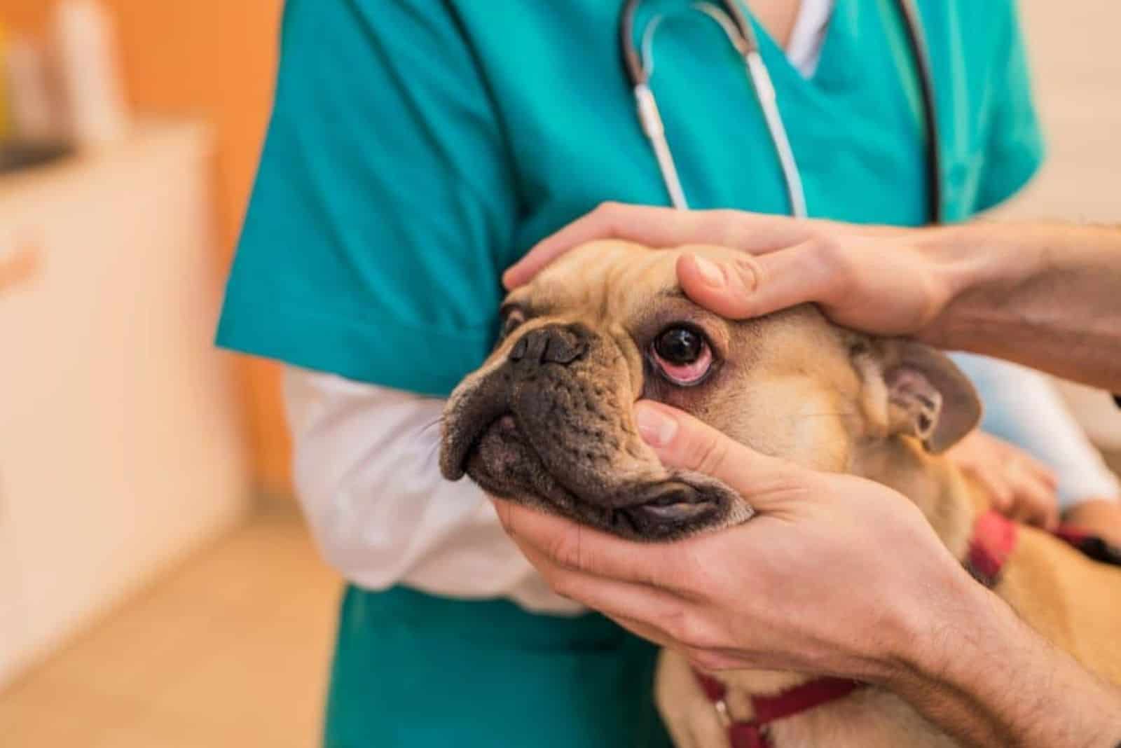
Dogs’ eyes are a crucial tool in the sense trifecta. I would say that the nose is the first line of environment analysis, followed by the ears and the eyes. Any problem concerning one of these elements will seriously impede your dog’s self-assuredness and interaction.
Some conditions that affect the eye are less serious than others, and most of them are manageable with medication. However, knowing how to interpret the symptoms on a very rudimentary level can help you preserve your dog’s quality of life.
We will go through all the common causes that beg the question, “Why are my dog’s eyes red?”. I believe it is time we put those worries aside and discuss eye conditions that cause redness.
Most Common Causes Of Red Eyes In Dogs
The list of conditions that cause eye redness in a dog is by no means short. However, some of the causes are more widespread than others. The simplest explanation is usually the right one, so eye injury is among the most common.
A foreign object stuck in your dog’s eye can cause a lot of damage, especially if the dog paws its eyes. All of these can be treated at home, but it is always best to call the vet and have a professional deal with it.
Conditions that are easily treatable by either medication or routine surgery include conjunctivitis (pink eye), cherry eye, and dry eye. To cover the entire spectrum of possible conditions and causes, let us have a closer look at each answer to “Why are my dog’s eyes red” in no particular order.
1. Keratoconjunctivitis Sicca (Dry Eye)
By the name of this condition, we can already presume what the problem is. However, there is a large number of things that can cause dry eye in a dog. A dry eye diagnosis means that your dog’s lacrimal gland and third eyelid (tear glands) are not producing enough tears.
The liquid that we know as tears is actually more complex than just “water”. It contains fats, water, and mucin. While only the water component is produced by the two aforementioned glands, it makes for the majority of its volume.
This water content is the portion that helps lubricate and reduce the chance of debris getting into the eye. The fat content of tears further lubricates the eye so that shutting your eyelids is comfortable and the eye moves smoothly.
Mucin contains extremely important antibacterial proteins, leukocytes, and mucus. These three components are arguably the most important segment that represents the first line of defense against germs.
In terms of causes, there are quite a few that can influence tear production and dry eye. The most common underlying problem is autoimmune disorders. The tear components are recognized as foreign bodies by the immune system and attacked by the white blood cells.
Endocrine problems (glands with internal secretion), such as Cushing’s, hypothyroidism, and diabetes mellitus, have dry eye as a symptom. Genetic predisposition to dry eye means that the condition is congenital (inherited).
If your dog is on medication therapy, the side effects of many meds can cause dry eyes, so discussing alternatives with your vet is of extreme importance. Injury, surgical procedures, infections, neurological disorders, and anesthesia are some other causes.
Symptoms Of Dry Eye
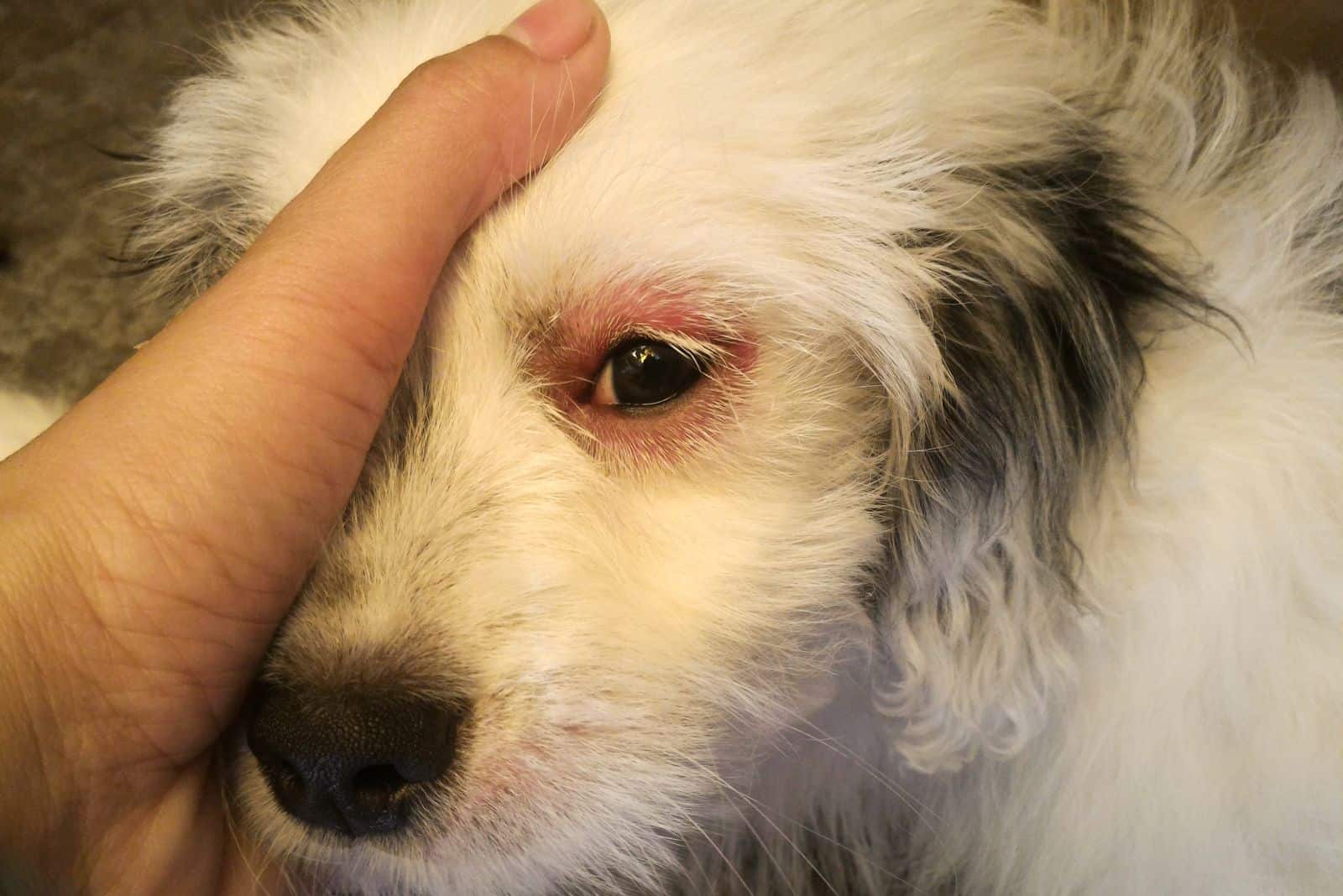
Since there are quite a few symptoms of KCS, you should easily notice them in your dog. Other than the red eyes, there can be swelling of the conjunctiva, eyelid, glands, or even less immediate tissue in the vicinity of the eye.
A dog that blinks or squints excessively or shows signs of temporarily degraded vision is likely to have dry eye.
Despite the lack of tear production, discharge that resembles mucus is a very common symptom of dry eye. Green or yellow discharge can indicate that there is a bacterial infection.
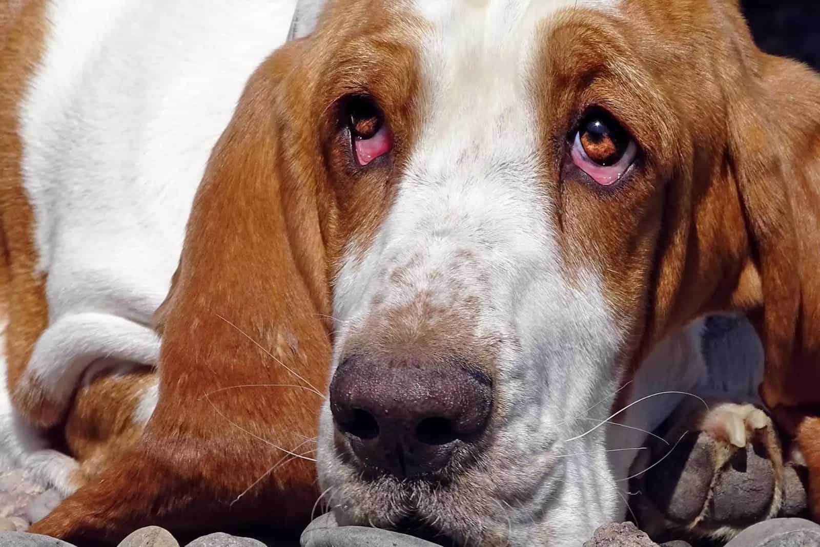
Eye redness is usually due to inflammation, but irregular vascularization is the main cause of that. Capillaries burst and create new blood vessels to try and replenish blood flow in the eye, changing the natural pigmentation of the cornea.
Diagnosis And Treatment
Your vet will most often use a test called the Schirmer Tear Test (STT) to diagnose dry eye. The process involves putting a thin filter paper on the inner side of the lower eyelid and waiting to see if adequate amounts of tears are produced.
Values of more than fifteen millimeters of tear liquid indicate that tear production is within normal range, while anything under ten millimeters means there is an issue with the tear glands.
Additionally, the DVM will do a fluorescein stain test (FST) to exclude potential corneal ulcers. Checking the ocular pressure is another way of narrowing it down to dry eye.
Common treatment options, depending on what the underlying cause of dry eye is, include ophthalmic cyclosporine (for autoimmune conditions) and tacrolimus (to make the eye increase tear production). This class of medicament is called lacrimostimulant.
If the lack of tear production cannot be reversed, then lacromimetics are used. This type of med is known as artificial tears. In the initial stages of dry eye, before the production resumes to normal, lacrimomimetics are used as a temporary replacement alongside lacrimostimulants.
For secondary eye infections that can occur with dry eye, oral antibiotics and ointments can be used to cure the infection and decrease inflammation in the eye. Surgery is only required for cases that do not improve with med therapy.
It is called parotid duct transposition and means that the surgeon will redirect the salivary gland’s duct from the mouth to supply saliva as a replacement liquid for tears.
2. Environmental Irritants (Allergies)
Similar to humans, dust, dust mites, mold, pollen, and other environmental irritants can cause your dog to have an allergic reaction that manifests itself as eye redness. Though typically rare in dogs, it is possible for allergies to affect the eye.
Medically, the condition is known as allergic conjunctivitis and is most often seen in dogs that have allergic dermatitis because of an already weakened immune system. If you know your dog has skin allergies that cause itching, or notice the latter, call a vet to schedule an appointment.
Brachycephalic breeds like the Pug and French Bulldog, Cocker Spaniels, Poodles, German Shepherds, and Golden Retrievers are more prone to dermatitis caused by seasonal allergies, so keep an eye out for symptoms.
Keep in mind that the chemical compounds used in perfumes, air fresheners, and certain detergents or fabric softeners can trigger an allergic reaction in your dog. Dog food can cause eye allergies but the eye in most cases remains asymptomatic for food allergies.
Symptoms Of Environmental Irritant-Induced Red Eye
Most symptoms related to red eyes overlap, so we are, once again, looking at excessive blinking, squinting, pawing, and eye discharge. Combined with itchiness, this will most likely point to an allergic reaction that affects the eyes too.
Diagnosis And Treatment
Performing conjunctival cytology is a method that has a mixed record of revealing cells that become active if an allergic reaction is active.
This means that additional testing and exclusion of other conditions are required to confirm environmental irritants as a factor. Taking a small piece of conjunctiva for a biopsy is a proven way to eliminate other options and pinpoint the cause.
Because it requires general anesthesia, it is a cumbersome procedure that has a recovery period, thus used only if needed. Instead, technological advancements in veterinary medicine allowed for a test called the conjunctival provocation test, which is faster and less intrusive.
Saline solutions can be used as an eye wash to clean your dog’s eyes one or two times per day. This will physically flush out any allergen that might be causing the reaction. Steroid-based eye drops are the treatment of choice for most dogs with eye allergies.
It is important not to use any medication without the veterinarian’s approval. You could do more damage than good if the cause is a condition that reacts negatively to owner-administered non-steroid anti-inflammatory meds.
For dogs that do not have a confirmed allergy diagnosis, allergy testing is mandatory to prevent further complications and immune system overload.
3. Uveitis
There are three types of uveitis in dogs, and they are named according to how many parts of the uvea are affected. Pan-uveitis means that the iris, ciliary body, and choroid are all inflamed.
The iris is the mechanism that adapts the circumference of the colored, circular “donut” in the eye. It controls the influx of light into the eye. The ciliary body is the glassy, watery body of the eye, and the choroid is the furthermost layer of the eye.
Because the front two parts (iris and ciliary body) are adjacent layers of the eye, uveitis that affects these two structures is called anterior (front of the eye) uveitis. Posterior uveitis means only the posterior part (choroid) has active inflammation.
Viral, fungal, and bacterial infections can all cause uveitis. Some well-known pathogenic diseases in dogs, such as distemper, leptospirosis, toxoplasmosis, blastomycosis, histoplasmosis, and others, can be causes of uveitis.
Diabetes and similar conditions that affect metabolism are commonly associated with this eye issue. Toxins introduced through the environment (typically chemical) can cause eye irritation that develops into uveitis.
Eye injury, high blood pressure, lens damage, or tumors are other possible causes that pretty much cover all of the possibilities.
Symptoms Of Uveitis
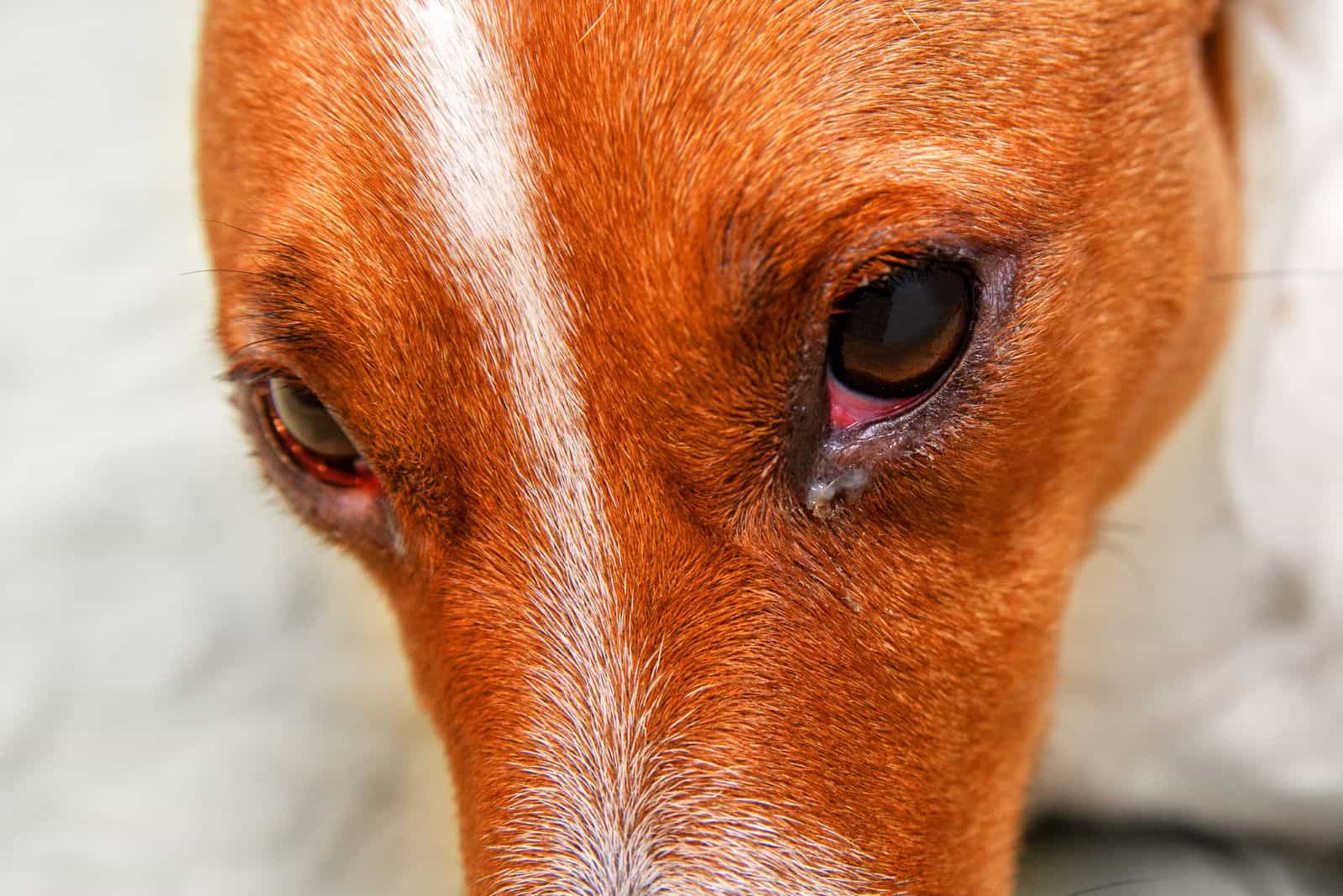
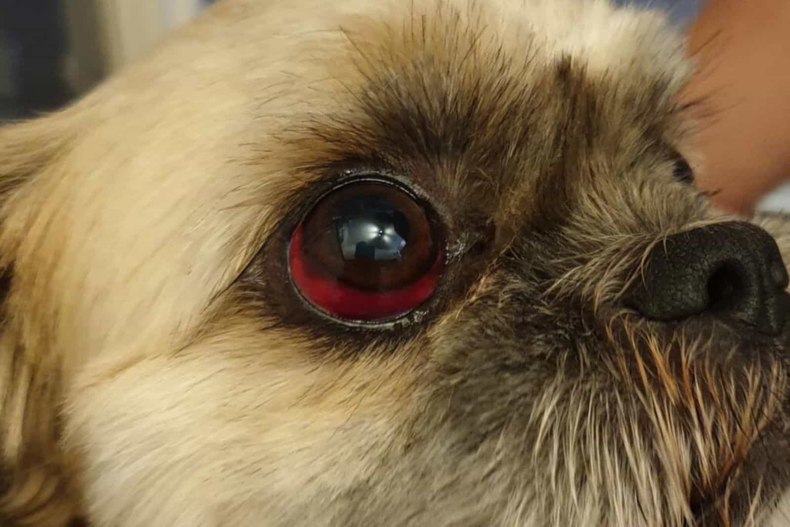
This condition can cause excessive tearing, unlike some of the other eye issues. The eyes become bloodshot, develop a bulging iris, iris constriction, cloudy eyes, and photophobia, pointing almost definitively at uveitis.
A permanent change in eye color is a possibility if the disease advances past the initial stage. Reversing the different coloration of the eye is not possible.
If acute uveitis progresses to a chronic state, your dog might develop cataracts, displacement of the lens, and, worst-case scenario, vision loss in older dogs. These symptoms are usually found in dogs that have severe cases of uveitis.
Diagnosis And Treatment
One of the most effective and painless tests is measuring intraocular pressure (IOP) to exclude glaucoma. With glaucoma, the eye pressure will be higher than if your dog has uveitis.
After that, a full physical examination is required to confirm the condition is not a symptom of some other eye problem or general illness. Testing the blood for viral, fungal, and bacterial infections will rule out pathogens as the cause.
Oral administration of corticosteroids and NSAIDs reduces inflammation and helps the dog cope with the pain. Oral medication can also include oral antibiotics in case of a bacterial infection.
Antibiotic eye drops are prescribed for usage during the day, while antibiotic ointments are preferred as a nighttime treatment for more lubrication while the dog’s eyes are shut.
If the damage is substantial due to a physical injury or the progression of uveitis has a high chance of creating future dog eye issues, then surgery is an option. Foreign object extraction or corneal tear reparation has a high success rate in terms of recurring eye problems.
4. Hyperthyroidism
The thyroid gland is essential in regulating a dog’s metabolism, body temperature, digestion, etc. Hyperthyroidism means that the gland is producing too much hormone (thyroxine and triiodothyronine).
In terms of a dog’s red eyes, hyperthyroidism can trigger hyperlipidemia, a condition that is characterized by excessive amounts of lipids in the blood. Hyperlipidemia does not directly cause redness in the eye, but the following conditions that develop from it do.
- lipemia retinalis
- lipemic aqueous humor
- corneal lipid dystrophy
- uveitis
All of these conditions can be at fault for a dog developing secondary eye problems, such as non-healing corneal erosion.
Symptoms Of Hyperthyroidism
While this condition does not necessarily display symptoms affecting the eye, hyperlipidemia is not that rare in dogs with an overly active thyroid.
Redness in the eye due to retinal bleeding and detachment is common with lipemia retinalis, while cloudy eyes are specific to lipemic aqueous humor due to free-floating lipids in the eyeball.
Corneal lipid dystrophy usually has both bloodshot and cloudy eyes as a symptom due to the fats putting pressure on the eyeball and causing blood vessels to rupture. Burst capillaries usually occur when corneal degeneration is the cause of corneal lipidosis.
Diagnosis And Treatment
Other than feeling the physical enlargement of the gland, a veterinary ophthalmologist will order a urinalysis, a full blood work panel, and a chemical blood profile.
If there are elevated concentrations of thyroxine (T4), it will be a clear sign of hyperthyroidism. A test called thyroid scintigraphy is used to determine whether the condition is congenital or because of thyroid carcinoma.
Treating hyperthyroidism is generally not problematic. Inhibition of thyroxine production is done via oral medication, even when the overproduction is caused by synthetic thyroid hormone therapy due to hypothyroidism.
Surgery treatment often includes removing the thyroid entirely, but radioactive iodine can be a less invasive way with a substantially easier recovery period.
5. Hyperadrenocorticism (Cushing’s Disease)
We have another condition that involves the excessive production of hormones. This time, the adrenal gland produces too much cortisol, leading to reduced efficiency in glucose management, tissue healing, and suppression of the immune system.
Medication that is based on steroids is often used to reduce inflammation in health conditions that affect dogs, so it is among the most common causes. Hyperadrenocorticism often causes Keratoconjunctivitis Sicca, leading to eye problems.
Symptoms Of Hyperadrenocorticism
Since the main eye issue caused by Cushing’s is KCS, redness in the eye, swelling of the eyeball and surrounding tissue (eyelids or corner of the eye), and colored discharge are common occurrences.
Other symptoms can include increased urination, excessive consumption of water, and increased appetite. Note that dogs with hyperadrenocorticism can develop stomach swelling without pain.
Diagnosis And Treatment
Detection and diagnosis of hyperadrenocorticism is a complex process. Three tests are performed to close in on a definitive conclusion. The adrenal gland is enlarged when it overproduces cortisol, so an ultrasound is used to analyze the size.
An ACTH stimulation test is performed by injecting a small dose of ACTH (hormone) that stimulates cortisol production. The amount of cortisol that is measured after the test will show if normal levels are raised.
The high-dose dexamethasone suppression (HDDS) test is usually done after a low-dose DS test was performed with a positive diagnosis for hyperadrenocorticism but did not clearly show which type.
If an adrenal or pituitary gland tumor is the cause behind the disease, radiation and chemotherapy can help shrink the tumor until it is small enough for surgical excision. For tumors that are caught early, immediate surgery, followed by radiation or chemo, is standard.
When steroidal medication is the reason for hyperadrenocorticism, discontinuation of therapy is the best available option. The reduction of hormone levels in most cases normalizes adrenal gland function.
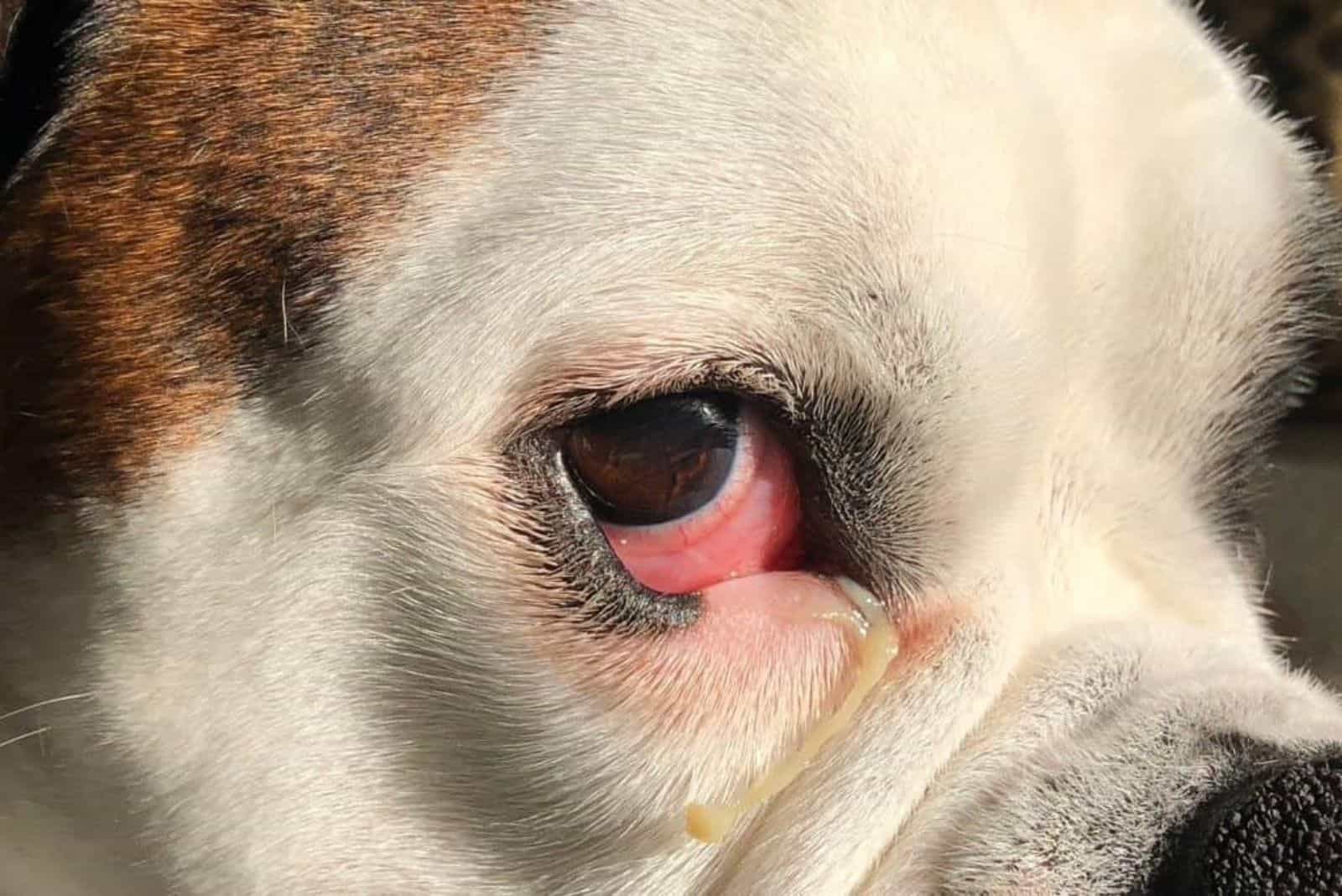
6. Cherry Eye
Dogs have a third eyelid that is located on the inner side of the lower eyelid. It is also called a nictating membrane, and its role is to protect the eye from debris and trauma.
As we saw at the beginning, this third eyelid also acts as a gland that produces tear components crucial for fighting germs. A prolapse of the nictating membrane happens when the connecting tissue that keeps it attached to the lower eyelid detaches.
Brachycephalic breeds are genetically predisposed to cherry eye due to the connection being malformed during the formative months of a dog. Boston terriers, Poodles, Cocker Spaniels, and some other breeds are also prone to it.
Symptoms Of Cherry Eye
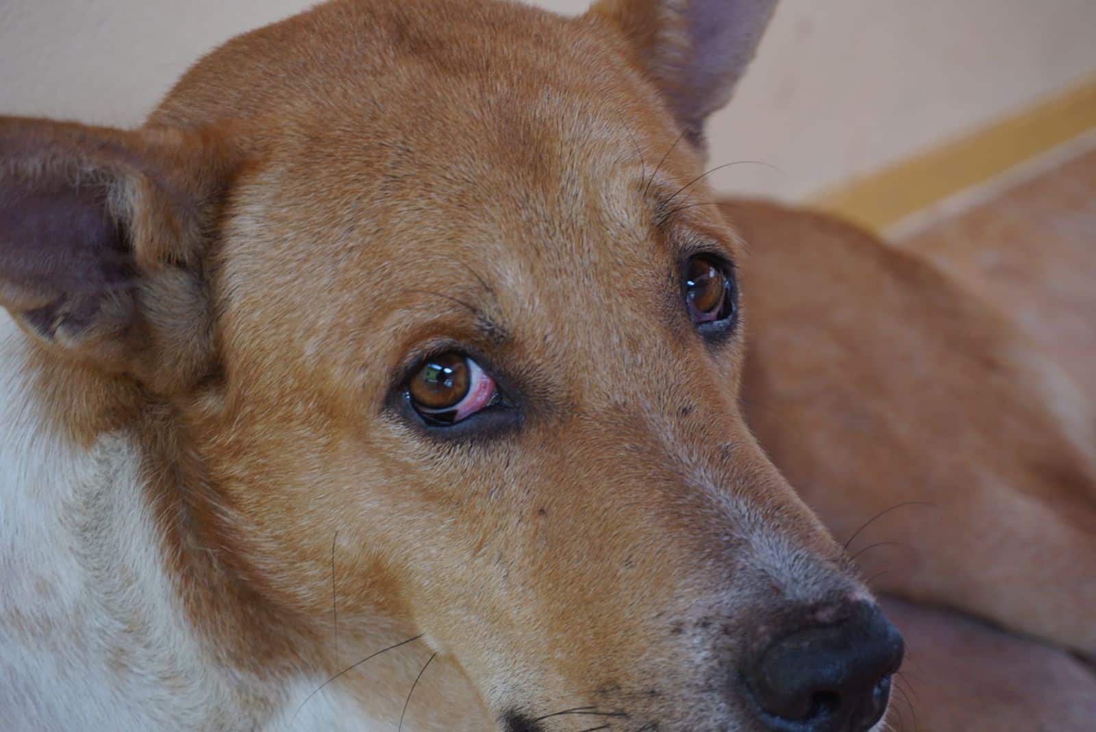
Although it is a normal part of a dog’s eye, the third eyelid will appear as a swollen mass of tissue. It will have a bright red color due to the inflammation and popped blood vessels. The redness will be present on the cornea because the third eyelid increases pressure on the eyeball.
While this is the only symptom, fluid build-up can be present, too. It can cause the appearance of excessive tearing, but it is simply an accumulation of mucus and tears that becomes trapped under the mass and moves in intervals.
Diagnosis And Treatment
It is crucial to take your dog to the vet as soon as you notice redness or swollen red tissue on the eye. The third eyelid has many vital roles in eye health, so getting the necessary surgery on time is of paramount importance.
The surgical procedure involves replacing the third eyelid. This is done by cutting the nictating membrane in several places and anchoring it with suturing knots to the lower eyelid. This way, the shape of the third eyelid and its anchor are restored to normal anatomy.
After the surgery, your vet will most likely give appropriate treatment in the form of NSAIDs to reduce the post-operative inflammation and antibiotic ointments or drops to curb any potential bacterial infection.
7. Glaucoma
Glaucomas in dogs come in two forms. One is chronic, and the other is acute. Chronic glaucoma is also known as open-angle glaucoma, and it inevitably leads to complete vision loss.
The sudden onset of glaucoma goes by the name of closed-angle glaucoma and is caused by an immediate increase in intraocular pressure. Three components of vision are damaged by a sudden spike in IOP — the optic nerve, the retina, and the optic disk.
The function of the iridocorneal angle (the duct that leads away excess fluids in the eye) becomes compromised, and liquid starts accumulating behind the eye. Due to fluid build-up, the increase in IOP forms glaucoma.
Symptoms Of Glaucoma
Glaucomas are divided into two groups based on their genesis. The first group includes genetic predisposition, while the second contains other eye problems like a luxating lens, cataracts, uveitis, and tumors.
Plenty of symptoms can point to glaucoma. The most commonly seen in affected dogs are non-responsiveness to light accompanied by pupil dilation, bulging bloodshot eyeballs, squinting, constant blinking, transparent discharge from the eye, pawing, and loss of coordination.
Severe cases of glaucoma will most likely have all of the above symptoms, but dogs with milder cases can display symptoms such as enlarged eyes, vein distention, cloudy eyes that have a blue sheen, and redness due to the capillaries bursting.
Diagnosis And Treatment
Using a tonometer, a device that measures IOP, the general vet will check the pressure in both eyes. If the pressure in either or both eyes is elevated, then your dog will be sent to a veterinary ophthalmologist for a gonioscopy.
This diagnostic process will allow the eye specialist to check the state of the iridocorneal angle. If results are inconclusive after gonioscopy, the ophthalmologist might do electroretinography or ultrasound.
Treating glaucoma is tough for severe cases where the optic nerve is heavily damaged. The main therapy will require lifelong administration of eye drops and pain meds. Alternatives include killing the cells that produce fluid in the eye by injection or cyclocryotherapy.
Ultimately, if the damage cannot be managed and puts the dog at risk for other health issues, then surgical eye removal is preferred over other forms of treatment.
Conclusion
It is never easy answering a health-related question because every dog has a unique set of circumstances. Questioning yourself, “Why are my dog’s eyes red?” can be stressful, as all dog owners like avoiding the veterinarian.
However, stuff happens that is out of our control, and your pooch might have to visit the dreaded DVM. In many cases, the conditions that cause red eye in dogs are nothing to worry about, and you will fix them with an appropriate treatment prescribed by your vet.
If your dog is unfortunate enough to have a more serious condition, the prognosis can still be good for most of the conditions in this article. Surely, your veterinarian will recommend the best possible treatment and explain everything to you.
No matter how insignificant the redness might be, my advice is you call the vet immediately or rush your dog to an animal hospital. It is better to be safe than sorry.
Read Next: Horner’s Syndrome In Dogs – An Eyelid Issue Or A Serious Problem?